Points to remember:
- Diastolic murmur extends throughout diastole
- Heard at 3rd left interspace
- Use diaphragm of stethoscope with firm pressure
- Have the patient hold expiratory breath
Discussion. There are two illustrations of Aortic Regurgitation on SAM II, the Student
Auscultation Manikin – Aortic Regurgitation and Acute Aortic Regurgitation.
Regurgitation occurs most commonly from damage to the valve leaflets, but also may
result from dilation of the aortic valve ring, damage to the ascending aorta, and rarely by
communications between the aorta and left ventricle.
Causes of Aortic Regurgitation in Adult Patients.
Severe Aortic Regurgitation. The most common causes of severe aortic regurgitation
in the adult are rheumatic heart disease and bacterial endocarditis. Antibiotics are
used to treat rheumatic fever so this cause is rapidly declining.
Mild Aortic Regurgitation. A common cause of mild aortic regurgitation is severe
hypertension. Aortic regurgitation occurs somewhat frequently with all types of
congenital aortic stenosis.
Musical AR murmurs often occur in patients with a perforated leaflet as in infective
endocarditis.
The length of the diastolic murmur may give an indication as to the severity of the
regurgitation. A long murmur may indicate moderate to severe AR. However, if the
murmur is very severe, the murmur may be harsh and surprisingly short for the degree
of regurgitation.
The diastolic murmur of AR sounds like an expiratory breath sound. This murmur occurs
immediately after the second heart sound. The second sound is generally either single
or very closely split with the A2-P2 interval being about.02 seconds. A split this close is
very hard for the listener to distinguish. The murmur is loudest in early diastole. There is
a short period of a crescendo and then it becomes decrescendo. The ear can
occasionally hear the crescendo, but essentially the murmur can be considered
decrescendo.
In his text on Clinical Heart Disease, W. Proctor Harvey, M.D. states that “The main, and
often the sole, diagnostic feature of aortic regurgitation is a diastolic murmur heard at the
aortic area or along the left sternal border”. Moderate to severe aortic regurgitation is
almost always associated with a systolic murmur (as well as a diastolic murmur) even in the absence of aortic stenosis.
Area of transmission.
When the murmur is loud, it may be heard over most of the precordium. In some cases,
when the left ventricle is enlarged, the murmur may be loudest along the left border of
the sternum, but is also well heard in an area above the apex in the anterior axillary
region. With pure aortic regurgitation, the aortic second sound is often accentuated. An
ejection sound may occur at the apex, but instead of being clicking in quality, it has the
same quality as the usual first sound and gives the impression of a split first sound.
The diastolic murmur of aortic regurgitation is usually loudest in the third left interspace,
but may occasionally maybe loudest in the second right interspace. When it is loudest in
the second right interspace, one must consider unusual conditions such as an aortic
aneurysm, a dissecting aneurysm, Marfan’s disease, etc.
Listening technique.
As noted in the lesson on Basic Cardiac Auscultation, the listener should begin by first
auscultating at the right upper sternal border (aortic area), followed by the left upper
sternal border (pulmonic area), and then proceed down the left sternal border (tricuspid
area) and finally the apex (mitral area). The stethoscope should be moved within the
above areas by inching with the area to determine the point of maximum intensity.
Palpate the carotid pulse to identify S1 and the onset of systole.
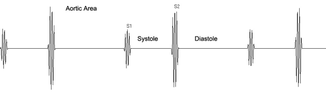
Aortic Area. There is a normal S1 followed by an ejection sound. S2 is followed by a
decrescendo diastolic murmur.
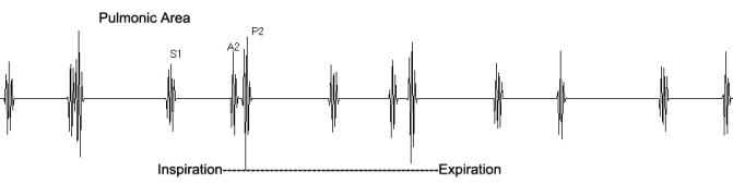
Pulmonic Area. At the pulmonic area, S1 is absent and P2 is loud due to moderate
pulmonary hypertension. The diastolic murmur is transmitted from the aortic area and is
grade II.
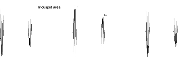
Tricuspid Area. Best heard. Aortic regurgitation murmurs are best heard with the
patient sitting upright, leaning forward with breath held in deep expiration. Use the
diaphragm of the stethoscope with firm pressure and listen along the third left sternal
border (Erb’s point). The stethoscope should be placed firm enough to leave an imprint
on the chest wall when removed. The firm pressure will help bring out a faint murmur.
Or the use of the E-Scope Electronic Stethoscope with the volume turned up will also be
of help in bringing out a faint murmur.
There will be a short systolic murmur followed by a decrescendo diastolic murmur that
occurs immediately after S2.
In younger patients, the murmur is often heard when they are in the recumbent position.
Best heard in the third or fourth left Intercostal Space.
Use diaphragm of stethoscope with firm pressure. The Patient should be leaning forward
and breathing out and holding the expiratory breath.
Pressure should be sufficient to cause an imprint of the stethoscope chest piece when removed.
Mitral Area (apex). An ejection sound may occur at the apex, but instead of being
clicking in quality, it has the same quality as the usual first sound and gives the
impression of a split first sound. The ejection sound may be followed by a decrescendo
Austin Flint murmur.
Maneuvers: 1. As described above, the patient should deeply exhale and hold his/her
breath while leaning forward. Listen with the diaphragm of the stethoscope placed at the
3rd left intercostal space with firm pressure. 2. Use a handgrip. As described by Jules
Constant, M.D., in his text Bedside Cardiology, page 386, “The elevation of systolic
blood pressure after 3 minutes of 33% maximum handgrip pressure is greater in patients
with AR and in normal subjects.” 4. A brisk upstroke in the radial pulse may be palpated.
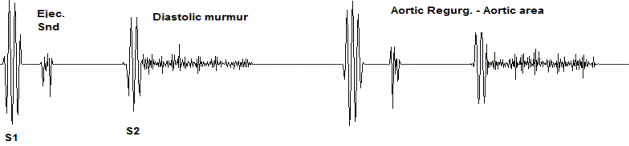
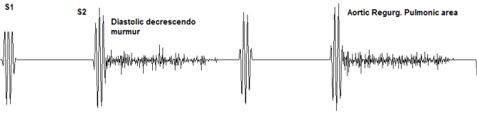
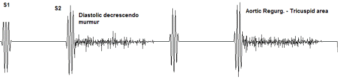
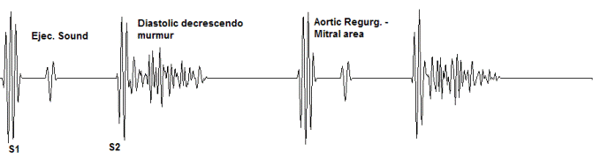
Figure 6 Aortic regurgitation. The Auscultatory Picture.
Mitral Regurgitation
Points to remember:
- Holosystolic murmur heard at the apex
Discussion. Review lesson on Mitral Regurgitation, Marked. The difference between
mild and marked mitral regurgitation from the standpoint of auscultation is that the
murmur of mild MR has a holosystolic murmur only in the mitral area while MR Marked
has a holosystolic murmur as well as an S3 and a diastolic murmur.
Mitral regurgitation, also known as mitral insufficiency, is a condition in which blood
partially returns to the left atrium from the left ventricle. It is due to one or more causes
such as integrity of the valve leaflets, proper size and function of the annulus, integrity of
all of the chordae tendineae, integrity and proper contraction of the papillary muscles
and proper function of the left ventricular muscle. Damage to any of these structures
may lead to regurgitation of blood from the left ventricle to the left atrium. There are
several causes such as rheumatic fever, prolapse of the mitral leaflets into the left atrium
during systole as in mitral valve prolapse, rupture of the chordae tendineae due to
infective endocarditis or non-penetrating chest trauma or left ventricular dilation.
One of the most common causes of mitral regurgitation is the prolapse of the mitral leaflets
into the left atrium during systole.
The classic sign of mitral regurgitation is the holosystolic murmur at the apex of the
heart. It is transmitted to the axilla and left back. A third heart sound is characteristic of
significant regurgitation and may be followed by a low-pitched mid-diastolic murmur.
The murmur of mitral regurgitation is pansystolic or holosystolic and is generated as
blood regurgitates from the left ventricle to the left atrium. The murmur is blowing,
medium-pitched and is best heard at the mitral area.
The most important characteristic of the systolic murmur of mitral regurgitation is that the
point of maximum intensity is at the apex (mitral area). It is, of course, systolic because
the blood regurgitates into the left atrium during ventricular contraction. When this
murmur is faint or moderately loud, it is high-pitched and blowing in character.
The typical systolic murmur is holosystolic, plateau in intensity and pitch and blowing in
quality. The murmur is usually transmitted loudly to the left axilla and left back.
Occasionally, the murmur may be transmitted to the upper sternal areas and even the
neck vessels that may cause confusion about the origin of the murmur.
In rheumatic disease, the first heart sound is usually diminished, but may be normal or
even increased in regurgitation of other etiologies. A third heart sound is characteristic
of significant regurgitation and may be followed by a low-pitched, mid-diastolic murmur.
A fourth heart sound is unusual. An opening snap, which is characteristic of mitral
stenosis, may be heard occasionally even in the absence of mitral stenosis.
In marked regurgitation, there is often a rather short, rumbling mid-diastolic murmur that
starts with the third heart sound. This occurs even in the absence of mitral stenosis and
is due to the marked increase in early diastolic inflow through the mitral valve. Mild MR
generally does not have an S3 or a diastolic murmur at the mitral areas as is found in
Marked MR.
It is important to distinguish the holosystolic murmur of mitral regurgitation from the
systolic murmur of aortic stenosis. Normally, you would expect to hear the murmur of
aortic stenosis at the aortic area. However, in severe aortic stenosis, it can also be
heard at the mitral area. The murmur of mitral regurgitation is holosystolic, that is, it
begins with S1 and continues through to S2. It is a high-pitched murmur and differs in
quality from that of aortic stenosis. Listen repeatedly to both mitral regurgitation and
then aortic stenosis until you can clearly tell the difference.
Listening technique.
As noted in the lesson on Basic Cardiac Auscultation, the listener should begin first by
auscultating at the right upper sternal border (aortic area) while palpating the carotid
pulse to determine S1 and the onset of systole. This should be followed by listening at
the left upper sternal border (pulmonic area), and then proceed down the left sternal
border (tricuspid area) and finally the apex (mitral area). The stethoscope should be
moved within the above areas by inching with the area to determine the point of
maximum intensity. Palpate the carotid pulse to identify S1 and the onset of systole.
Maneuvers:
A sustained handgrip will increase the loudness of the murmur of mitral regurgitation.
Best heard. The murmur of mitral regurgitation is best heard at the apex (mitral area).
Aortic area. The first and second sounds (S1 and S2) are heard with S2 being
somewhat dominant.
Pulmonary area. At the pulmonic area, S1 is heard with A2 and P2 being split at about
0.06 seconds.
Tricuspid area. At the tricuspid area, S1 is normal and dominant over S2.
Mitral area. (Best heard) At the mitral area, S1 and S2 are heard. There is a
holosystolic murmur between S1 and S2.
In patients with marked mitral regurgitation, there if often an S3 following S2 by about
msec. and a middiastolic murmur.
Mitral Area
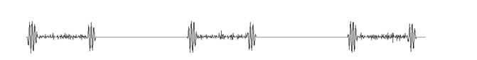
Figure 7 Mitral regurgitation. The Auscultatory Picture.