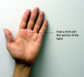
A Thrill is a tremor or vibration the can be felt with a grade 4, 5 or 6 murmur. In aortic
stenosis, if a systolic thrill is present, it will be felt in the right upper or upper middle part
 of the chest. The direction of the thrill is toward the right neck; the direction of a thrill of
pulmonic stenosis is toward the left neck.
When feeling a Thrill, there are two factors to consider – the location and the rhythm. A
thrill may be systolic, diastolic or both. The location is also a factor in making a
diagnosis.
If a systolic thrill is felt in the upper right sternal border close to the sternum or near
the aortic area, may be aortic stenosis. A diagnosis cannot be made by the thrill alone,
but only in combination with other physical signs.
A thrill maybe systolic and diastolic, systolic only or diastolic only. A diastolic thrill at
the apex may be associated with mitral regurgitation or mitral stenosis. A systolic and
diastolic thrill may be a sign of a pericardial friction rub. This can be confirmed by
auscultation.
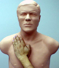
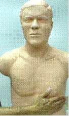
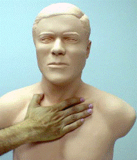
Thrill at aortic area Thrill at apical area Thrill at upper chest
Normal Heart Sounds
Points to remember:
- Second sound is split (S2 = A2 + P2)
- Splitting of S2 occurs on inspiration, closes on expiration
- Split S2 heard in the pulmonic area
- P2 closes later than A2
- Increased P2 indicates pulmonary hypertension
Aortic area. The first and second heart sounds (S1 and S2) are normal with the second
heart sound being dominant in both the aortic and pulmonic areas.
Pulmonic area. Identify the A2 and P2 components of S2. Listen to the Pulmonic Area
(second left intercostal space -2LICS) and identify S2. S2 is the loudest in this area. The
first protuberance below the suprasternal notch is the “ angle of Louis ”. It is the point
where the sternum and the second costal cartilage are joined. Below this hump is the
second intercostal space. The angle of Louis can be helpful in identifying the second
right or left intercostal space.
Listen to normal heart sounds at the pulmonic area. You will hear normal physiologic
splitting on inspiration with a normal or slightly split S2 on expiration. Splitting is normally
between 0.03 to 0.07 seconds. When the second sound is split, the aortic component is
termed A2 and the pulmonic component is termed P2. A loud P2 may occur in pulmonary
hypertension. Generally, an S3 is not loud enough to be heard in this area.
Tricuspid Area. Listen at the tricuspid area (mid-left sternal border). S1 and S2 are
normal. Although not shown in this example, S1 may be slightly split. A split S1 is
difficult to hear, as the split is very close. The first heart sound is dominant in both the
tricuspid and mitral areas. At the left sternal border, S1 may be split because you begin
to hear the tricuspid closure. S1 splits into the mitral (M1) and tricuspid (T1) sounds. You
may hear an S3 also in younger patients. An S3 is not shown in this example.
Mitral area. First, identify S1, which is the onset of systole. Palpate the carotid pulse and listen for
the first heart sound at the mitral area. The key element in cardiac auscultation is the
identification of S1. If systole and diastole are not properly identified, then all the sounds
will be incorrect.
Place the stethoscope in the Mitral Area (apex) where S1 is louder than S2. Palpate
the carotid pulse. The pulse will coincide with S1 and indicates the onset of systole.
Diastole is about 1/3 longer than systole at a normal heart rate of 75-80 bpm. As the heart
rate increases, diastole becomes shorter until, at about 105-110 bpm, it is about the
same length as systole. At this rate, the onset of systole is difficult to distinguish by
sound alone. Palpating the carotid pulse will assist the listener in determining the onset
of systole and, hence, recognizing S1.


Figure 4 Normal heart sounds. The Auscultatory Picture.
Aortic Stenosis Mild
Points to remember:
- Systolic ejection diamond-shaped murmur that ends before S2. The later


 of the chest. The direction of the thrill is toward the right neck; the direction of a thrill of


