Figures 4 and 5 show the primary areas for
listening during an auscultation examination. Area 1 in Figure 4 is the aortic area – also
called the second right sternal border (2RSB). Area 2 is the pulmonic area also called
the second left sternal border (2LSB).
Area 3 is the tricuspid area (4LSB) and area 4 is the mitral or apical area or the fifth left
intercostal space (5LICS, also called the Midclavicular line or MCL). As you progress,
you will find that additional areas are necessary during cardiac auscultation such as
Erb’s point which is the 3rd left intercostal space (3LICS), the Left Ventricular Area and
the Right Ventricular Area.
The Left Ventricular Area is located around the apex and extends to the anterior axillary
line.
The Right Ventricular Area is located in both the right and left lower part of the sternum
and includes the 3rd and 4th intercostal spaces
These auscultatory areas are now usually designated by surface anatomical location as
shown in the above paragraph such as (4LSB ). For purposes of this lesson, we will
use the traditional designations – aortic, pulmonic, tricuspid and mitral.
Whenever a sound is best heard in one of these auscultatory areas, it is most likely to
have been produced in the region of the valve for which it is named. The dotted lines in
Figure 4 shows the region where the point of maximum intensity may be found. The best
location for listening can vary from individual to individual.
When moving your stethoscope within these anatomical areas to find the point of
maximum intensity (PMI), move the stethoscope slowly over the area until the optimum
location for the sound is heard. This is known as “inching”.
For a thorough examination, the patient should be in a sitting, lying or left lateral
recumbent position. For an adequate examination, the recumbent position alone cannot
be used and listening to the heart with the patient only in a sitting position is absolutely
inadequate. Typical diastolic murmurs of good intensity that are heard when the patient
is in the recumbent position may NOT be heard at all when the patient is sitting. In the
recumbent position, the patient’s arms must not be held over the head, since this will
elevate the rib cage and decrease the intensity of the heart sound.
Occasionally, it is advantageous to listen with the patient in the prone position; this may
be true in patients with deep chests or those who may have a friction rub. This position
may be awkward when the patient is in bed or on the examining table and supporting
himself/herself on the elbows. It is much more satisfactory to have the patient stand up
and lean over with his/her elbows on an examining table. Muscle noise is at a minimum
in this position and the patient and examiner are much more comfortable. An important
additional advantage of this position is that the patient can be made to exercise by
touching his toes several times before he assumes the position.
Patient Positioning.
Another important method for finding the optimum sound is
patient positioning. If the patient is seated upright, have him or her lean forward. This
moves the heart nearer the solid chest wall improving the transmission of sound. If the
patient is in bed, have them turn onto the left side (left lateral position) to produce the
same effect.
Listening technique.
Listening to heart sounds. According to Thornton and Karnath1 and most
cardiologists, a systematic approach should be followed when listening to the heart. The
listener should begin by first auscultating at the right upper sternal border (aortic area),
followed by the left upper sternal border (pulmonic area), and then proceed down the left
sternal border (tricuspid area) and finally to the apex (mitral area). The stethoscope
should be moved within the above areas by a process known as “inching”. This allows
the listener to locate the point of maximum intensity
Frequency of heart sounds
Knowledge of frequency is very helpful to the understanding of cardiac auscultation.
Heart sounds range from 20 to 650Hz. Above 650Hz, there is little diagnostic
information. The first and second heart sounds are in the frequency range of 70-120Hz.
Murmurs are generally between 150-400Hz. The third and fourth heart sounds are very
low in frequency – about 40-60Hz. If you suspect an S3, you must listen very carefully as
the frequency is very low. An S3 is generally best heard at the mitral area. An S4 can be
heard in either the mitral area or along the left sternal border. In general, you will hear
an S4 in the mitral area. There will be the occasional patient where an S4 is heard along
the left sternal border.
Murmurs
What is a murmur? A heart murmur is an extra or unusual sound heard during a
heartbeat. Murmurs range from very faint to very loud. They sometimes sound like a
whooshing or swishing noise. Normal heart-beats make a "lub-DUB" sound. This is the
sound of the heart valves closing as blood moves through the heart.
There are two types of heart murmurs: innocent (harmless) and abnormal. People who
have innocent heart murmurs have normal hearts. They usually have no other signs or
symptoms of heart problems. Innocent murmurs are common in healthy children. Many,
if not most, children will have heart murmurs heard by their doctors at some time in their
lives.
People who have abnormal murmurs may have other signs or symptoms of heart
problems. Most abnormal murmurs in children are due to congenital heart defects.
These are heart defects that are present at birth. In adults, abnormal murmurs are most
often due to heart valve problems caused by infection, disease, or aging.
A heart murmur isn't a disease, and most murmurs are harmless. Innocent murmurs
don't cause symptoms or require you to limit physical activity. Although an innocent
murmur may be a lifelong condition, if the heart is normal it is probably that treatment will
not be needed.
The outlook and treatment for abnormal heart murmurs depends on the type and the
severity of the heart problem causing them.
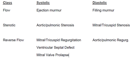
Classification of Murmurs
Most murmurs can be correlated to the underlying pathology. It is usually possible to
say that a murmur is the murmur of mitral regurgitation or aortic stenosis or mitral
stenosis. Each of these murmurs has certain characteristics by which it can be
recognized. Their characteristics, in order of importance, are:
- Gross timing – that is, systolic or diastolic
- Point of maximum intensity
- Pitch and quality
- Finer timing
It is not difficult to determine whether a murmur is systolic or diastolic when the rate is
slow, but it may be difficult with more rapid rates. Again, palpation of the carotid pulse
will assist in locating the first heart sound that is the onset of systole in adult patients.
Murmurs are generally classified as follows:
Grading of Murmurs
Murmurs are graded for loudness from 1 to 6 with 6 being the loudest. This grading
system can be in Roman numerals or Arabic. Generally, this grading is expressed as
2/6 or II/VI
Palpation of a Thrill