MR Marked differs from Mild MR in that it has a diastolic murmur at the apex as
well as a systolic murmur.
Mitral Stenosis
Points to remember:
- Listen with diaphragm
- Has Opening snap after S2
- Accentuated S1
- Rarer forms of MS have a presystolic murmur (called presystolic
Accentuation)
Discussion. Valvulitis associated with one of the rheumatic fever syndromes is thought
to be the cause of mitral stenosis in essentially all patients with this disorder.
Mitral stenosis is an obstruction to flow across the mitral orifice that may be caused by
adhesions between the two mitral leaflets, loss of mobility of the leaflets and possibly
fibrosis and rigidity of the chordae tendineae and papillary muscles. It can be caused by
valvulitis associated with one of the rheumatic fever syndromes and is thought to be the
cause of mitral stenosis in essentially all patients with this disorder. The primary cause of
mitral stenosis is rheumatic fever and, until recently with an influx of immigrants, is heard
infrequently in the US and other developed countries.
The classic features of mitral stenosis are: 1) an accentuated first heart sound, 2) an
opening snap of the mitral valve, 3) a mid-diastolic murmur and 4) a presystolic murmur
(called a presystolic accentuation).
Auscultation remains the principle method of detecting mitral stenosis. The diastolic
rumble at the apex is almost diagnostic in itself. This is especially true if there is
presystolic increase in pitch and intensity of the rumble and if the first sound is loud and
an opening snap is detected.
The murmurs of mitral stenosis are diastolic and maximum at the apex (mitral area).
Even when loud at the apex, they may not be well transmitted anywhere else in the
precordium. Their finer timing is such that they are considered mid-diastolic and
presystolic. An accentuated first heart sound and an opening snap of the mitral valve
are very characteristic of mitral stenosis and may be the earliest findings. The
accentuated first heart sound results from fibrotic changes in the mitral valve and
chordae tendineae and possibly form a higher closing pressure.
The opening snap of mitral stenosis occurs between 0.06 and 0.10 seconds (60 to 100
msec.) after the aortic second sound and the more severe the mitral stenosis, the closer
it occurs to the aortic second sound.
The earliest murmur in mitral stenosis is usually a sort of tumbling mid-diastolic murmur that
is produced as blood flows through the narrowed mitral valve during the phase of rapid
filling of the ventricle. This murmur is best heard at the apex (mitral area) with the
patient in the left lateral position and with the bell applied lightly to the chest wall. If the
bell is applied too firmly and the murmur is not very loud, the murmur may not be
detected.
Auscultation remains the principle method of detecting mitral stenosis. In pure mitral
stenosis, the diagnosis is not difficult and is made by auscultation. When there is
multiple valvular diseases, the severity of the mitral stenosis may be more difficult to
determine. The echocardiogram provides a good estimation of the degree of the
obstruction. Cardiac catheterization may be necessary in some complex cases. In any
patient with pulmonary hypertension, mitral stenosis should be carefully considered and
either excluded or established.
Listening technique.
As noted in the lesson on Basic Cardiac Auscultation, the listener should begin first by
auscultating at the right upper sternal border (aortic area) while palpating the carotid
pulse to determine S1 and the onset of systole. This should be followed by listening at
the left upper sternal border (pulmonic area), and then proceed down the left sternal
border (tricuspid area) and finally the apex (mitral area). The stethoscope should be
moved within the above areas by inching with the area to determine the point of
maximum intensity. Palpate the carotid pulse to identify S1 and the onset of systole.
Maneuvers:
A sustained handgrip will increase the loudness of the murmur of mitral stenosis.
Where to Listen:
Best heard. The murmur of mitral stenosis is best heard at the apex (mitral area) using
the bell of the stethoscope.
Aortic area. The first heart sound is increased in intensity to mitral
Pulmonary area. The first heart sound is increased in intensity and louder than the
second heart sound. In some cases, the second sound may be split with P2 being
increased due to pulmonary hypertension. In this example, however, S2 is not split.
Tricuspid area. At the tricuspid area, S1 is accentuated and dominant over S2. An
opening snap is present after S2. In some cases there may be a grade II/VI systolic
murmur due to tricuspid regurgitation from papillary muscle dysfunction secondary to
right ventricular dilation due to severe pulmonary hypertension. The example shown
with SAM II does not have a murmur at the tricuspid area.
Mitral area. At the mitral area, S1 is accentuated and an opening snap is present
approximately 70 msec. (0.07 seconds) from S2. There is also a mid-diastolic murmur
and a presystolic accentuation (murmur).
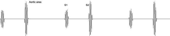
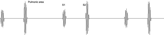
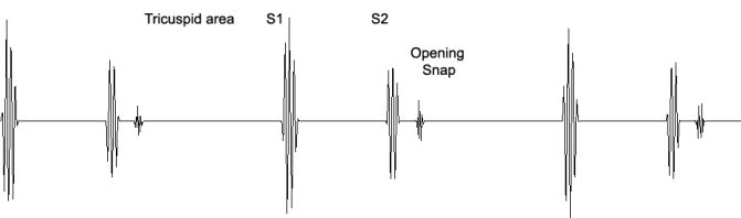

Figure 7 Mitral stenosis. The Auscultatory Picture.
Ventricular Septal Defect (VSD)
Points to remember:
- Heard in tricuspid area
- No systolic wave in the neck
- Harsh, blowing murmur ends before S2
- Heard in children
- Heard in adults with anterior infarction
Discussion. Ventricular septal defect (VSD) is a common congenital heart lesion
encountered in infants and children. Many of these defects will close spontaneously
with age. Ventricular septal defects are classified as small, medium or large with the
small normally closing during the first few years of life.
A holosystolic murmur, grade 3-4, is heard along the mid and lower left sternal border
and at the mitral area (apex). The murmur of VSD usually extends throughout systole,
that is, it is
holosystolic. It may be of equal intensity throughout systole, but often shows some
peaking during systole and may be diamond-shaped. The point of maximum intensity is
usually in the third or fourth left intercostal space to the left of the sternum. When the
murmur is faint or moderately loud, it tends to be high-pitched with mild harshness and is
somewhat harsher than the murmur of mitral regurgitation.
The difference in the quality of faint and moderately loud murmurs of VSD and mitral
regurgitation is not great and the point of maximum intensity is more valuable for
differentiation.
Occasionally, small ventricular septal defects have a diamond-shaped murmur that ends
before the second sound. A third heart sound at the apex is quite common and is
probably the result of increased flow through the mitral valve.
A middiastolic murmur can be heard at the apex in large ventricular septal defects (not
illustrated in this example) and, as in the case of patent ductus arteriosus, is due to
increased flow through the mitral valve.
Maneuvers: A sustained handgrip increases the murmur loudness.
Listening technique. As noted in the lesson on Basic Cardiac Auscultation, the listener
should begin by first auscultating at the right upper sternal border (aortic area), followed
by the left upper sternal border (pulmonic area), and then proceed down the left sternal
border (tricuspid area) and finally the apex (mitral area). The stethoscope should be
moved within the above areas by inching with the area to determine the point of
maximum intensity.
In small children, the femoral artery is an adequate time reference for determining the
onset of systole and may be more practical than the carotid in short necks.
Best heard. The murmur of ventricular septal defect is best heard along the lower left
sternal border at the 3rd left interspace.
Aortic area. There is a normal S1 and S2 with S1 being slightly dominant.
Pulmonic area. There is a normal S1 and S2 with a short, medium frequency, grade 1
systolic murmur due to normal turbulence of flow across the pulmonary outflow tract.
The murmur is diamond-shaped and softer than the murmur in the tricuspid area.
Tricuspid area. S1 and S2 are normal and there are high frequency holosystolic
murmur1 due to flow across the ventricular septal defect
Mitral area. S1 and S2 are normal. There is a holosystolic murmur that has been
transmitted from the tricuspid area. There is an S3 indicating that the shunt is medium or
large.
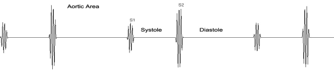
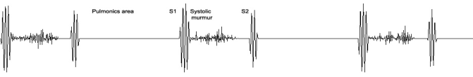

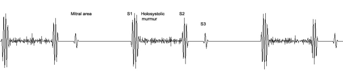
Figure 8 Ventricular Septal Defect. The Auscultatory Picture.
Atrial Septal Defect (ASD)
Points to remember:
- Fixed splitting of the second sound is the classic feature of ASD
- Spitting of S2 is in both inspiration and expiration
- A systolic murmur usually accompanies split S2
Discussion. One of the key characteristics of the atrial septal defect (ASD) is fixed splittingof the second sound in the second left interspace. The second sound is almost always split during both inspiration and expiration and there is little or no effect of inspiration on this splitting.
Additionally, ASDs are almost always associated with a systolic murmur in the second
left interspace. The murmur is of medium pitch and is rarely as harsh or loud as that of a
large ventricular septal defect (VSD). The ASD murmur is due to a marked increase of
blood flow through the pulmonary valve and relative pulmonary stenosis as a result of
enlargement of the right ventricle and pulmonary artery. The murmur and the fixed
splitting can be heard at the pulmonic area of SAM II, the Student Auscultation Manikin.
The length of the systolic murmur is variable depending upon the size of the atrial septal
defect.
In patients with more marked shunts, a rumbling diastolic murmur is heard along the left
border of the sternum. This can be heard in the tricuspid area of SAM. This is a
tricuspid flow murmur and resembles, in many ways, the murmur of mitral stenosis
except that it occurs more medially.
At times, the first heart sound is increased in intensity because of the accentuation of the
tricuspid component. This increased first sound is heard along the left border of the
sternum and in the mesocardiac area. If the pulmonic component of the split second
sound is mistaken for an opening snap of the mitral valve, it is easy to mistake an atrial
septal defect that has a mid-diastolic murmur and an accentuated first sound for mitral
stenosis.
Aside from the fact that the phenomena are heard more medially than would be
expected in mitral stenosis, differentiation between ASD and mitral stenosis depends, in the good part, on recognition of the fact that one is dealing with a split second sound with
fixed splitting rather than an opening snap of the mitral valve. The expected effect of
respiration of the second sound in mitral stenosis and the lack of effect in atrial septal
defect can be recognized by listening carefully in the second left intersperse.
In mitral stenosis, there is a single second sound in expiration followed by an opening
snap. In inspiration, the pulmonic second sound moves into the space between the
aortic second sound and the opening snap and there are three sounds: the aortic
sound, the pulmonic second sound and the opening snap.
Maneuvers: Valsalva maneuver.
The behavior of the second heart sound after a Valsalva maneuver is quite different in
normal persons from that of patients with ASD and this difference has diagnostic
significance. During a Valsalva maneuver, the right side of the heart is suddenly flooded
with a large amount of blood so that the right ventricular systole is prolonged and there is
marked splitting of the second sound by as much as 0.10 seconds. Within the next four
to six beats, this sudden load has been transferred from the right side of the heart to the
left side of the heart and now the left ventricular contraction is prolonged and right
ventricular contraction shortened. The aortic and pulmonic sounds are superimposed
and only a single sound is heard.
The occurrence of a single sound after several beats is an important observation and
sometimes, when one cannot decide whether there is splitting in expiration, the Valsalva
maneuver can be used to see whether the split disappears. In atrial septal defects,
there is essentially a common atrium and a large amount of blood that reaches the
atria at the end of the Valsalva maneuver affects filling of both ventricles so that the
splitting which was present before the maneuver continues unchanged after the
maneuver and the sound do not close into a single sound.
Listening technique.
As noted in the lesson on Basic Auscultation, the listener should begin by first
auscultating at the right upper sternal border (aortic area), followed by the left upper
sternal border (pulmonic area), and then proceed down the left sternal border (tricuspid
area) and finally the apex (mitral area). The stethoscope should be moved within the
above areas by inching with the area to determine the point of maximum intensity.
In small children, the femoral pulse is an adequate time reference for determining the
onset of systole and may be more practical than the carotid in short necks.
Best heard. The murmur of atrial septal defect is best heard at the lower left sternal
border.
Aortic area. S1 and S2 are normal and no murmurs are heard.
Pulmonic area. S2 is widely split at 0.07 seconds and does not move with respiration.
Tricuspid area. There is a mid-diastolic murmur that is due to increased flow across a
normal tricuspid valve as shunted blood recirculates from the left to right atrium and into the
right ventricle.
Mitral area. S1 and S2 are normal and no murmurs are heard.

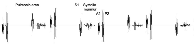
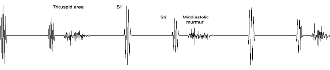
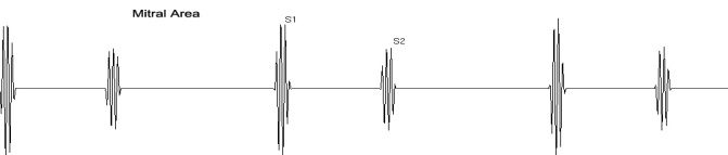
Figure 8 Atrial Septal Defect. The Auscultatory Picture.
Pulmonary Stenosis
Points to remember:
- Systolic
- Ejection click
- Chest and axillae (often loudest in axillae)
- Newborn infants (especially premature)
Discussion.
There is a difference between pulmonic stenosis with an intact septum and pulmonic
stenosis with a non-intact septum, the latter being essentially the same as a ventricular
septal defect. The example in this sound is with an intact septum.
This condition is associated with a harsh, systolic murmur of maximum intensity in the
second intercostal space, an abnormally split sound, a pulmonary ejection click and
often a right atrial or fourth heart sound. The murmur is the most obvious finding.
Because it takes longer for the right ventricle to empty through the stenosed pulmonary
valve, the right ventricular systole is prolonged over the left ventricular systole and there is an abnormally wide splitting of the second sound. If the pulmonary stenosis is mild, the pulmonic second sound may be of fairly normal intensity, it is delayed in both expiration and inspiration with the inspiratory delay being somewhat greater. With moderate stenosis, the pulmonic second sound becomes faint and in severe stenosis, it may not be heard. The murmur is harsh like that of aortic stenosis and is also often diamond-shaped. The more severe the stenosis, the greater the prolongation of right ventricular systole and the longer the murmur. Also, the peak of the murmur is earlier in systole with mild stenosis and in severe stenosis; there is a longer murmur with a late peak.
Note that with the long murmur and the long delay in the pulmonic second sound, the
murmur extends through the aortic second sound so that the aortic second sound is not
heard at the second left intercostal space, but may be heard in the second right
intercostal space.
A pulmonary ejection sound is nearly always present and is best heard in the second
intercostal space. It varies with respiration and is louder in expiration. The ejection
sound may be masked by the loud murmur and will often be heard better in the third left
intercostal space when the murmur is not as loud.
Infundibular stenosis is really pulmonary stenosis without the ejection sound. The length
of the murmur is a function of the severity of both pulmonary stenosis and infundibular
stenosis. Mild is shorter and more severe is longer.
Listening technique. As noted in the lesson on Basic Auscultation, the listener should begin by first auscultating at the right upper sternal border (aortic area), followed by the left upper sternal border (pulmonic area), then proceed down the left sternal border (tricuspid area) and finally to the apex (mitral area). The stethoscope should be moved within the above areas by inching within the area to determine the point of maximum intensity.
Aortic area. There is a normal S1 and S2 with an S2 being dominant. A systolic murmur
of medium frequency appears in late systole.
Pulmonic area. S1 is followed by an ejection click that increases with expiration and
decreases with inspiration. Breath sounds are not illustrated here in order to allow the
student to concentrate on the murmur. There is an abnormally split second sound (P2)
along with the pulmonary ejection click. The murmur of pulmonic stenosis is usually
present from infancy, but may not be detected until adulthood. The murmur is maximal in
the second left interspace and radiates to the left side of the neck and sometimes the left
posterior shoulder. A thrill is common, usually in the second left intercostal space.
Tricuspid area. There is a transmitted systolic murmur. No S4 is heard.
Mitral area. There is a grade I systolic murmur transmitted.