The remainder of the thoracic spinal nerves, T3 through T12, do little recombining. They form the intercostal nerves, so named because they run between the ribs. For points of reference, the 7th intercostal nerve terminates at the lower end of the sternum, also known as the xyphoid process. The 10th intercostal nerve terminates at the umbilicus, or the belly button.
The somatic nervous system is that part of the peripheral nervous system associated with the voluntary control of body movements through the action of skeletal muscles, and also reception of external stimuli. The somatic nervous system consists of afferent fibers that receive information from external sources, and efferent fibers that are responsible for muscle contraction. The somatic system includes the pathways from the skin and skeletal muscles to the Central Nervous System. It is also described as involved with activities that involve consciousness.
The basic route of the efferent somatic nervous system includes a two neuron sequence. The first is the upper motor neuron, whose cell body is located in the precentral gyrus (Brodman Area 4) of the brain. It receives stimuli from this area to control skeletal (voluntary) muscle. The upper motor neuron carries this stimulus down the corticospinal tract and synapses in the ventral horn of the spinal cord with the alpha motor neuron, a lower motor neuron. The upper motor neuron releases acetylcholine from its axon terminal knobs and these are received by nicotinic receptors on the alpha motor neuron. The alpha motor neurons cell body sends the stimulus down its axon via the ventral root of the spinal cord and proceeds to its neuromuscular junction of its skeletal muscle. There, it releases acetylcholine from its axon terminal knobs to the muscles nicotinic receptors, resulting in stimulus to contract the muscle.
The Sympathetic and Parasympathetic Systems
The sympathetic nervous system activates what is often termed the fight or flight response, as it is most active under sudden stressful circumstances (such as being attacked). This response is also known as sympathetico-adrenal response of the body, as the pre-ganglionic sympathetic fibers that end in the adrenal medulla (but also all other sympathetic fibers) secrete acetylcholine, which activates the secretion of adrenaline (epinephrine) and to a lesser extent noradrenaline (norepinephrine) from it. Therefore, this response that acts primarily on the cardiovascular system is mediated directly via impulses transmitted through the sympathetic nervous system and indirectly via catecholamines secreted from the adrenal medulla.
Western science typically looks at the SNS as an automatic regulation system, that is, one that operates without the intervention of conscious thought. Some evolutionary theorists suggest that the sympathetic nervous system operated in early organisms to maintain survival (Origins of Consciousness, Robert Ornstein; et al.), as the sympathetic nervous system is responsible for priming the body for action. One example of this priming is in the moments before waking, in which sympathetic outflow spontaneously increases in preparation for action.
The parasympathetic nervous system is part of the autonomic nervous system. Sometimes called the rest and digest system or feed and breed. The parasympathetic system conserves energy as it slows the heart rate, increases intestinal and gland activity, and relaxes sphincter muscles in the gastrointestinal tract.
After high stress situations (ie: fighting for your life) the parasympathetic nervous system has a backlash reaction that balances out the reaction of the sympathetic nervous system. For example, the increase in heart rate that comes along with a sympathetic reaction will result in an abnormally slow heart rate during a parasympathetic reaction.
Organization
Sympathetic nerves originate inside the vertebral column, toward the middle of the spinal cord in the intermediolateral cell column (or lateral horn), beginning at the first thoracic segment of the spinal cord and extending into the second or third lumbar segments. Because its cells begin in the thoracic and lumbar regions of the spinal cord, the SNS is said to have a thoracolumbar outflow. Axons of these nerves leave the spinal cord in the ventral branches (rami) of the spinal nerves, and then separate out as 'white rami' (so called from the shiny white sheaths of myelin around each axon) which connect to two chain ganglia extending alongside the vertebral column on the left and right. These elongated ganglia are also known as paravertebral ganglia or sympathetic trunks. In these hubs, connections (synapses) are made which then distribute the nerves to major organs, glands, and other parts of the body.
In order to reach the target organs and glands, the axons must travel long distances in the body, and, to accomplish this, many axons link up with the axon of a second cell. The ends of the axons do not make direct contact, but rather link across a space, the synapse.
In the SNS and other components of the peripheral nervous system, these synapses are made at sites called ganglia. The cell that sends its fiber is called a preganglionic cell, while the cell whose fiber leaves the ganglion is called a postganglionic cell. As mentioned previously, the preganglionic cells of the SNS are located between the first thoracic segment and the second or third lumbar segments of the spinal cord. Postganglionic cells have their cell bodies in the ganglia and send their axons to target organs or glands.
The ganglia include not just the sympathetic trunks but also the superior cervical ganglion (which sends sympathetic nerve fibers to the head), and the celiac and mesenteric ganglia (which send sympathetic fibers to the gut).
Messages travel through the SNS in a bidirectional flow. Efferent messages can trigger changes in different parts of the body simultaneously. For example, the sympathetic nervous system can accelerate heart rate; widen bronchial passages; decrease motility (movement) of the large intestine; constrict blood vessels; increase peristalsis in the esophagus; cause pupil dilation, piloerection (goose bumps) and perspiration (sweating); and raise blood pressure. Afferent messages carry sensations such as heat, cold, or pain.
The first synapse (in the sympathetic chain) is mediated by nicotinic receptors physiologically activated by acetylcholine, and the target synapse is mediated by adrenergic receptors physiologically activated by either noradrenaline or adrenaline. An exception is with sweat glands which receive sympathetic innervation but have muscarinic acetylcholine receptors which are normally characteristic of PNS. Another exception is with certain deep muscle blood vessels, which have acetylcholine receptors and which dilate (rather than constrict) with an increase in sympathetic tone. The sympathetic system cell bodies are located on the spinal cord excluding the cranial and sacral regions, specifically the thoracolumbar region (T1-L3). The preganglonic neurons exit from the vertebral column and synapse with the postganglonic neurons in the sympathetic trunk.
The parasympathetic nervous system is one of three divisions of the autonomic nervous system. Sometimes called the rest and digest system, the parasympathetic system conserves energy as it slows the heart rate, increases intestinal and gland activity, and relaxes sphincter muscles in the gastrointestinal tract.
Receptors
The parasympathetic nervous system uses only acetylcholine (ACh) as its neurotransmitter. The ACh acts on two types of receptors, the muscarinic and nicotinic cholinergic receptors. Most transmissions occur in two stages: When stimulated, the preganglionic nerve releases ACh at the ganglion, which acts on nicotinic receptors of the postganglionic nerve. The postganglionic nerve then releases ACh to stimulate the muscarinic receptors of the target organ.
The three main types of muscarinic receptors that are well characterised are:
· The M1 muscarinic receptors are located in the neural system.
· The M2 muscarinic receptors are located in the heart, and act to bring the heart back to normal after the actions of the sympathetic nervous system: slowing down the heart rate, reducing contractile forces of the atrial cardiac muscle, and reducing conduction velocity of the atrioventricular node (AV node). Note, they have no effect on the contractile forces of the ventricular muscle.
· The M3 muscarinic receptors are located at many places in the body, such as the smooth muscles of the blood vessels, as well as the lungs, which means that they cause vasoconstriction and bronchoconstriction. They are also in the smooth muscles of the gastrointestinal tract (GIT), which help in increasing intestinal motility and dilating sphincters. The M3 receptors are also located in many glands that help to stimulate secretion in salivary glands and other glands of the body.
·
The Nerve cell
· The watershed of all studies of the nervous system was an observation made in 1889 by Spanish scientist Santiago Ramón y Cajal, who reported that the nervous system is composed of individual units that are structurally independent of one another and whose internal contents do not come into direct contact. According to his hypothesis, now known as the neuron theory, each nerve cell communicates with others through contiguity rather than continuity. That is, communication between adjacent but separate cells must take place across the space and barriers separating them. It has since been proved that Cajal’s theory is not universally true, but his central idea—that communication in the nervous system is largely communication between independent nerve cells—has remained an accurate guiding principle for all further study.
· There are two basic cell types within the nervous system: neurons and neuroglial cells.
The neuron
· In the human brain there are an estimated 85 billion to 200 billion neurons. Each neuron has its own identity, expressed by its interactions with other neurons and by its secretions; each also has its own function, depending on its intrinsic properties and location as well as its inputs from other select groups of neurons, its capacity to integrate those inputs, and its ability to transmit the information to another select group of neurons.
· With few exceptions, most neurons consist of three distinct regions, as shown in the diagram: (1) the cell body, or soma; (2) the nerve fibre, or axon; and (3) the receiving processes, or dendrites.
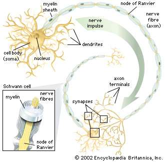
Anatomy of a nerve cellStructural features of a motor neuron include the cell body, nerve fibres, and dendrites.Encyclopædia Britannica, Inc.
Soma
Plasma membrane
· The neuron is bound by a plasma membrane, a structure so thin that its fine detail can be revealed only by high-resolution electron microscopy. About half of the membrane is the lipid bilayer, two sheets of mainly phospholipids with a space between. One end of a phospholipid molecule is hydrophilic, or water attaching, and the other end is hydrophobic, or water repelling. The bilayer structure results when the hydrophilic ends of the phospholipid molecules in each sheet turn toward the watery mediums of both the cell interior and the extracellular environment, while the hydrophobic ends of the molecules turn in toward the space between the sheets. These lipid layers are not rigid structures; the loosely bonded phospholipid molecules can move laterally across the surfaces of the membrane, and the interior is in a highly liquid state.
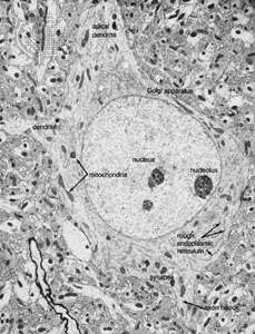
A neuron from the visual cortex of a ratThe centre of the field is occupied by the cell body, or soma, of the neuron. Most of the cell body is occupied by the nucleus, which contains a nucleolus. The double membrane of the nucleus is surrounded by cytoplasm, containing elements of the Golgi apparatus lying at the base of the apical dendrite. Mitochondria can be seen dispersed in the cytoplasm, which also contains the rough endoplasmic reticulum. Another dendrite is seen to the side, and the axon hillock is shown at the initial segment of the emerging axon. A synapse impinges onto the neuron close to the axon hillock.Courtesy of Alan Peters
· Embedded within the lipid bilayer are proteins, which also float in the liquid environment of the membrane. These include glycoproteins containing polysaccharide chains, which function, along with other carbohydrates, as adhesion sites and recognition sites for attachment and chemical interaction with other neurons. The proteins provide another basic and crucial function: those which penetrate the membrane can exist in more than one conformational state, or molecular shape, forming channels that allow ions to pass between the extracellular fluid and the cytoplasm, or internal contents of the cell. In other conformational states, they can block the passage of ions. This action is the fundamental mechanism that determines the excitability and pattern of electrical activity of the neuron.
· A complex system of proteinaceous intracellular filaments is linked to the membrane proteins. This cytoskeleton includes thin neurofilaments containing actin, thick neurofilaments similar to myosin, and microtubules composed of tubulin. The filaments are probably involved with movement and translocation of the membrane proteins, while microtubules may anchor the proteins to the cytoplasm.
Nucleus
· Each neuron contains a nucleus defining the location of the soma. The nucleus is surrounded by a double membrane, called the nuclear envelope, that fuses at intervals to form pores allowing molecular communication with the cytoplasm. Within the nucleus are the chromosomes, the genetic material of the cell, through which the nucleus controls the synthesis of proteins and the growth and differentiation of the cell into its final form. Proteins synthesized in the neuron include enzymes, receptors, hormones, and structural proteins for the cytoskeleton.
Organelles
· The endoplasmic reticulum (ER) is a widely spread membrane system within the neuron that is continuous with the nuclear envelope. It consists of series of tubules, flattened sacs called cisternae, and membrane-bound spheres called vesicles. There are two types of ER. The rough endoplasmic reticulum (RER) has rows of knobs called ribosomeson its surface. Ribosomes synthesize proteins that, for the most part, are transported out of the cell. The RER is found only in the soma. The smooth endoplasmic reticulum (SER) consists of a network of tubules in the soma that connects the RER with the Golgi apparatus. The tubules can also enter the axon at its initial segment and extend to the axon terminals.
· The Golgi apparatus is a complex of flattened cisternae arranged in closely packed rows. Located close to and around the nucleus, it receives proteins synthesized in the RER and transferred to it via the SER. At the Golgi apparatus, the proteins are attached to carbohydrates. The glycoproteins so formed are packaged into vesicles that leave the complex to be incorporated into the cell membrane.
Axon
· The axon arises from the soma at a region called the axon hillock, or initial segment. This is the region where the plasma membrane generates nerve impulses; the axon conducts these impulses away from the soma or dendrites toward other neurons. Large axons acquire an insulating myelin sheath and are known as myelinated, or medullated, fibres. Myelin is composed of 80 percent lipid and 20 percent protein; cholesterol is one of the major lipids, along with variable amounts of cerebrosides and phospholipids. Concentric layers of these lipids separated by thin layers of protein give rise to a high-resistance, low-capacitance electrical insulator interrupted at intervals by gaps called nodes of Ranvier, where the nerve membrane is exposed to the external environment. In the central nervous system the myelin sheath is formed from glial cells called oligodendrocytes, and in peripheral nerves it is formed from Schwann cells (see below The neuroglia).
· While the axon mainly conducts nerve impulses from the soma to the terminal, the terminal itself secretes chemical substances called neurotransmitters. The synthesis of these substances can occur in the terminal itself, but the synthesizing enzymes are formed by ribosomes in the soma and must be transported down the axon to the terminal. This process is known as axoplasmic flow; it occurs in both directions along the axon and may be facilitated by microtubules.
· At the terminal of the axon, and sometimes along its length, are specialized structures that form junctions with other neurons and with muscle cells. These junctions are called synapses. Presynaptic terminals, when seen by light microscope, look like small knobs and contain many organelles. The most numerous of these are synaptic vesicles, which, filled with neurotransmitters, are often clumped in areas of the terminal membrane that appear to be thickened. The thickened areas are called presynaptic dense projections, or active zones.
· The presynaptic terminal is unmyelinated and is separated from the neuron or muscle cell onto which it impinges by a gap called the synaptic cleft, across which neurotransmitters diffuse when released from the vesicles. In nerve-muscle junctions the synaptic cleft contains a structure called the basal lamina, which holds an enzyme that destroys neurotransmitters and thus regulates the amount that reaches the postsynaptic receptors on the receiving cell. Most knowledge of postsynaptic neurotransmitter receptors comes from studies of the receptor on muscle cells. This receptor, called the end plate, is a glycoprotein composed of five subunits. Other neurotransmitter receptors do not have the same structure, but they are all proteins and probably have subunits with a central channel that is activated by the neurotransmitter.
· While the chemically mediated synapse described above forms the majority of synapses in vertebrate nervous systems, there are other types of synapses in vertebrate brains and, in especially great numbers, in invertebrate and fish nervous systems. At these synapses there is no synaptic gap; instead, there are gap junctions, direct channels between neurons that establish a continuity between the cytoplasm of adjacent cells and a structural symmetry between the pre- and postsynaptic sites. Rapid neuronal communication at these junctions is probably electrical in nature. (For further discussion, see below Transmission at the synapse.)
Dendrites
· Besides the axon, neurons have other branches called dendrites that are usually shorter than axons and are unmyelinated. Dendrites are thought to form receiving surfaces for synaptic input from other neurons. In many dendrites these surfaces are provided by specialized structures called dendritic spines, which, by providing discrete regions for the reception of nerve impulses, isolate changes in electrical current from the main dendritic trunk.
· The traditional view of dendritic function presumes that only axons conduct nerve impulses and only dendrites receive them, but dendrites can form synapses with dendrites and axons and even somata can receive impulses. Indeed, some neurons have no axon; in these cases nervous transmission is carried out by the dendrites.
The neuroglia
· Neurons form a minority of the cells in the nervous system. Exceeding them in number by at least 10 to 1 are neuroglial cells, which exist in the nervous systems of invertebrates as well as vertebrates. Neuroglia can be distinguished from neurons by their lack of axons and by the presence of only one type of process. In addition, they do not form synapses, and they retain the ability to divide throughout their life span. While neurons and neuroglia lie in close apposition to one another, there are no direct junctional specializations, such as gap junctions, between the two types. Gap junctions do exist between neuroglial cells.
·
·
Types of neuroglia
· Apart from conventional histological and electron-microscopic techniques, immunologic techniques are used to identify different neuroglial cell types. By staining the cells with antibodies that bind to specific protein constituents of different neuroglia, neurologists have been able to discern two (in some opinions, three) main groups of neuroglia: (1) astrocytes, subdivided into fibrous and protoplasmic types; (2) oligodendrocytes, subdivided into interfascicular and perineuronal types; and sometimes (3) microglia.
· Fibrous astrocytes are prevalent among myelinated nerve fibres in the white matter of the central nervous system. Organelles seen in the somata of neurons are also seen in astrocytes, but they appear to be much sparser. These cells are characterized by the presence of numerous fibrils in their cytoplasm. The main processes exit the cell in a radial direction (hence the name astrocyte, meaning “star-shaped cell”), forming expansions and end feet at the surfaces of vascular capillaries.
· Unlike fibrous astrocytes, protoplasmic astrocytes occur in the gray matter of the central nervous system. They have fewer fibrils within their cytoplasm, and cytoplasmic organelles are sparse, so that the somata are shaped by surrounding neurons and fibres. The processes of protoplasmic astrocytes also make contact with capillaries.
· Oligodendrocytes have few cytoplasmic fibrils but a well-developed Golgi apparatus. They can be distinguished from astrocytes by the greater density of both the cytoplasm and the nucleus, the absence of fibrils and of glycogen in the cytoplasm, and large numbers of microtubules in the processes. Interfascicular oligodendrocytes are aligned in rows between the nerve fibres of the white matter of the central nervous system. In gray matter, perineuronal oligodendrocytesare located in close proximity to the somata of neurons. In the peripheralnervous system, neuroglia that are equivalent to oligodendrocytes are called Schwann cells.
· Microglial cells are small cells with dark cytoplasm and a dark nucleus. It is uncertain whether they are merely damaged neuroglial cells or occur as a separate group in living tissue.
Neuroglial functions
· The term neuroglia means “nerve glue,” and these cells were originally thought to be structural supports for neurons. This is still thought to be plausible, but other functions of the neuroglia are now generally accepted. Oligodendrocytes and Schwann cells produce the myelinsheath around neuronal axons. Some constituent of the axonal surface stimulates Schwann cell proliferation; the type of axon determines whether there is loose or tight myelination of the axon. In tight myelination a glial cell wraps itself like a rolled sheet around a length of axon until the fibre is covered by several layers. Between segments of myelin wrapping are exposed sections called nodes of Ranvier, which are important in the transmission of nerve impulses. Myelinated nerve fibres are found only in vertebrates, leading biologists to conclude that they are an adaptation to transmission over relatively long distances.
· Another well-defined role of neuroglial cells is the repair of the central nervous system following injury. Astrocytes divide after injury to the nervous system and occupy the spaces left by injured neurons. The role of oligodendrocytes after injury is unclear, but they may proliferate and form myelin sheaths.
· When neurons of the peripheral nervous system are severed, they undergo a process of degeneration followed by regeneration; fibres regenerate in such a way that they return to their original target sites. Schwann cells that remain after nerve degeneration apparently determine the route. This route direction is also performed by astrocytes during development of the central nervous system. In the developing cerebral cortex and cerebellum of primates, astrocytes project long processes to certain locations, and neurons migrate along these processes to arrive at their final locations. Thus, neuronal organization is brought about to some extent by the neuroglia.
· Astrocytes are also thought to have high-affinity uptake systems for neurotransmitters such as glutamate and gamma-aminobutyric acid (GABA). This function is important in the modulation of synaptic transmission. Uptake systems tend to terminate neurotransmitter action at the synapses and may also act as storage systems for neurotransmitters when they are needed. For instance, when motor nerves are severed, nerve terminals degenerate and their original sites are occupied by Schwann cells. The synthesis of neurotransmitters by neurons apparently also requires the presence of neuroglial cells in the vicinity.
· Finally, the environment surrounding neurons in the brain consists of a network of very narrow extracellular clefts. In 1907 Italian biologist Emilio Lugaro suggested that neuroglial cells exchange substances with the extracellular fluid and in this way exert control on the neuronal environment. It has since been shown that glucose, amino acids, and ions—all of which influence neuronal function—are exchanged between the extracellular space and neuroglial cells. After high levels of neuronal activity, for instance, neuroglial cells can take up and spatially buffer potassium ions and thus maintain normal neuronal function.
Sources:
https://en.wikipedia.org/wiki/Nervous_system
http://www.innerbody.com/image/nervov.html
https://www.ncbi.nlm.nih.gov/pubmedhealth/PMHT0025454/
https://en.wikibooks.org/wiki/Human_Physiology/The_Nervous_System
Unit 11
Urinary system

T opic Urinary System
Grammar Infinitive and its Syntactic Functions Revision – ing forms
Vocabulary Nervous System, Noun suffix – age, Adjective suffix – able, Verb suffix - en Revision Noun suffixes – um, - us Adjective suffixes– al, - ive(-ative, -itive), - ary Verb suffix - ate
Reading ‘Urinary System’
Listening ‘Human Urinary System’ (video)
Speaking Pair work (Structure and Functions of US), Group Work

Getting started
45. Study the facts relating to the Urinary System and answer the questions:
ü What other facts do you know about kidneys and urinary system?
ü What parts does the urinary system consist of?
ü Think of three functions of thekidneys and urinary system. What is the main function of the kidneys?
ü What branch of medicine deals withkidneys and urinary system?
ü Who is a specialist, working in the field of urology?
Vocabulary for help ing you to cope with texts and tasks:
 Word building:
Noun Suffix – age
Adjective Suffix - able
Verb suffix - en
See Unite 5 for the adjective suffix – al
See Unite 7 the verb suffix – ate,adjective suffix – ary
See Unite 9 for theadjective suffix – ive(-ative, -itive)
Noun suffix – age typically forms mass or abstract nouns from various parts of speechoccurring originally in loanwords from French (voyage, courage) and is productive in English with the meanings “aggregate”(coinage; peerage; trackage), “process” (coverage; breakage), “the outcome of” as either “the fact of” or “the physical effect or remains of” (seepage; wreckage; spoilage), “place of living or business”(parsonage; brokerage), “social standing or relationship” (bondage; marriage; patronage), and “quantity, measure, or charge” (footage; shortage; tonnage; towage).
Adjective suffix – able a suffix meaning “capable of, susceptible of, fit for, tending to, given to,” associated in meaning with the word able, occurring in loanwords from Latin (laudable); used in English as a highly productive suffix to form adjectives by addition to stems of any origin (teachable; photographable).
Verb suffix – en isformerly used to form transitive and intransitive verbs from adjectives (fasten; harden;sweeten), or from nouns (heighten;lengthen; strengthen).
1. Choose verbs (nouns, adjectives) form the words given below and translate them into Russian:
Urinary, renal, eliminate, regulate, chemical, primary, stable, abdominal, locate, physical, damage, medial, extensive, functional, central, environmental, individual, vertebral, connective, longitudinal, transitional, storage, flatten, mucosal, internal, involuntary, external, urethral, voluntary, active, epithelial, coverage.
2. Read and translate the anatomical terms of Latin-Greek origin. Pay your attention to the pronunciation of the words:
Renal[`rJn(q)l], ureter [juq`rJtq], urethra [juq`rJTrq], volume [`vOljHm], urea[juq`rJq], uric[`juqrIk], balance[`bxlqns], enzyme [`enzaIm], medial[`mJdIql], cavity[`kxvqtI], parenchyma[pq`reNkImq], sinus[`saInqs], glomerulus[glO`mFr(j)Vlqs], nephron[`nFfrOn], tubule[`t(j)HbjHl], peristalsis[,perI`stxlsIs], circular[`sE:kjVlq], epithelium[,epI`TJlIqm], submucosa[,sAbmjV`kqVsq], muscularis[`mAskjVlqrIs], sphincter[`sfIN(k)tq], synthesis[`sInTqsIs] (pl. syntheses [`sInTqsJs]), urothelium[,jVqrq(V)`TJlIqm]
Useful vocabulary: 
excretorysystem[eks`krJtqrI `sIstqm]– выделительнаясистема
urinarysystem[`juqrIn(q)rI`sIstqm]– мочевыделительнаясистема
renal[`rJn(q)l] - почечный
urinarytract[`juqrIn(q)rI trxkt] – мочевыделительныепути
kidney[`kIdnI] - почка
ureter[juq`rJtq]- мочеточник
urinarybladder[`juqrIn(q)rI `blxdq] – мочевойпузырь
urethra[juq`rJTrq]– уретра, мочеиспускательный канал
toeliminate[I`lImIneIt] - устранять, удалять
wasteproducts[weIst `prOdAkts] - продуктыжизнедеятельности
volume[`vOljHm] - объём, уровень
bloodvolume [blAd `vOljHm] – объёмциркулирующейкрови
todissolve[dI`zOlv] - растворять, разжижать
urea[juq`rJq]- мочевина
uricacid[`juqrIk] – мочеваякислота
damage[`dxmIG]- повреждение
indentation[,Inden`teIS(q)n] – углубление, лунка
renalsinus[`rJn(q)l `saInqs]– почечнаяпазуха
renalcortex[`rJn(q)lkLteks] - корковоевеществопочки
renalmedulla[`rJn(q)lme`dAlq] - мозговоевеществопочки
renaltubule[`rJn(q)l `t(j)HbjHl] (kidneytubule) – почечныйканалец
renalpelvis[`rJn(q)l `pelvIs] - почечнаялоханка
blood supply[blAd sq`plaI] - кровоснабжение
to leave [lJv] (left, left) - оставлять, покидать
glomerulus[glO`mFr(j)Vlqs] - клубочек
ureterovesicalvalve[juq,rJtqV`vFsIk(q)l vxlv]– сфинктермочеточника
toprevent[prI`vent]- предотвращать, предупреждать, проводить профилактику
toflow[flqu] - течь, струится
toempty[`emptI] - опорожнять, опустошать
coat[kqut]– оболочка, покров, слой
transitionalepithelium[trxn`zIS(q)nql,epI`TJlIqm] – переходныйэпителий
tostore[stL]– хранить, накапливать
toexpel[Ik`spel]–выгонять, исключать
urine[`juqrIn] - моча
toexpand[Ik`spxnd]- растягиваться, расширяться
urination [,juqrI`neIS(q)n] - мочеиспускание
voiding[`vOIdIN] – опорожнение, опустошение
sphincter[`sfIN(k)tq] - сфинктер
tocontribute (to) [kqn`trIbjHt]– способствовать, вноситьвклад
tocoverwith[`kAvq] – покрывать, закрывать
toflatten[`flxt(q)n]– делать плоским, выравнивать