Intraperitoneal parts of the GI tract are covered with serosa. These include most of the stomach, first part of the duodenum, all of the small intestine, caecum and appendix, transverse colon, sigmoid colon and rectum. In these sections of the gut there is clear boundary between the gut and the surrounding tissue. These parts of the tract have a mesentery.
Retroperitoneal parts are covered with adventitia. They blend into the surrounding tissue and are fixed in position. For example, the retroperitoneal section of the duodenum usually passes through the transpyloric plane. These include the esophagus, pylorus of the stomach, distal duodenum, ascending colon, descending colon and anal canal. In addition, the oral cavity has adventitia.
Basic structure
The gastrointestinal tract is a muscular tube lined by a special layer of cells, called epithelium. The contents of the tube are considered external to the body and are in continuity with the outside world at the mouth and the anus. Although each section of the tract has specialised functions, the entire tract has a similar basic structure with regional variations.

The wall is divided into four layers as follows:
Mucosa
The innermost layer of the digestive tract has specialised epithelial cells supported by an underlying connective tissue layer called the lamina propria. The lamina propria contains blood vessels, nerves, lymphoid tissue and glands that support the mucosa. Depending on its function, the epithelium may be simple (a single layer) or stratified (multiple layers).
Areas such as the mouth and oesophagus are covered by a stratified squamous (flat) epithelium so they can survive the wear and tear of passing food. Simple columnar (tall) or glandular epithelium lines the stomach and intestines to aid secretion and absorption. The inner lining is constantly shed and replaced, making it one of the most rapidly dividing areas of the body! Beneath the lamina propria is the muscularis mucosa. This comprises layers of smooth muscle which can contract to change the shape of the lumen.
Submucosa
The submucosa surrounds the muscularis mucosa and consists of fat, fibrous connective tissue and larger vessels and nerves. At its outer margin there is a specialized nerve plexus called the submucosal plexus or Meissner plexus. This supplies the mucosa and submucosa.
Muscularis externa
This smooth muscle layer has inner circular and outer longitudinal layers of muscle fibres separated by the myenteric plexus or Auerbach plexus. Neural innervations control the contraction of these muscles and hence the mechanical breakdown and peristalsis of the food within the lumen.
Serosa/mesentery
The outer layer of the GIT is formed by fat and another layer of epithelial cells called mesothelium.
Liver
The liver is not only the largest gland in the body but also the most complex in function.
Gross anatomy
The liver lies under the lower right rib cage and occupies much of the upper right quadrant of the abdomen, with a portion extending into the upper left quadrant. The organ weighs from 1.2 to 1.6 kg (2.6 to 3.5 pounds) and is somewhat larger in men than in women. Its greatest horizontal measurement ranges from 20 to 22 cm (approximately 8 inches); vertically, it extends 15 to 18 cm, and in thickness it ranges from 10 to 13 cm. The liver is divided into two unequal lobes: a large right lobe and a smaller left lobe. The left lobe is separated on its anterior (frontal) surface by the dense falciform (sickle-shaped) ligament that connects the liver to the undersurface of the diaphragm. On the inferior surface of the liver, the right and left lobes are separated by a groove containing the teres ligament, which runs to the navel. Two small lobes, the caudate and the quadrate, occupy a portion of the inferior surface of the right lobe. The entire liver, except for a small portion that abuts the right leaf of the diaphragm, is enveloped in a capsule of tissue that is continuous with the parietal peritoneum that lines the abdominopelvic walls and diaphragm.

Anterior and posterior views of the liver.
The major blood vessels enter the liver on its inferior surface in a centrally placed groove called the porta hepatis, which anatomically separates the quadrate and caudate lobes. The liver has two sources of blood supply: fully oxygenated blood from the hepatic artery, which is a major branch of the celiac axis (the main artery that crosses the abdomen) after its emergence from the abdominal aorta; and partially oxygenated blood from the large portal vein, which in turn receives all venous blood from the spleen, pancreas, gallbladder, lower esophagus, and the remainder of the gastrointestinal tract, including the stomach, small intestine, large intestine, and upper portion of the rectum. The portal vein is formed by the juncture of the splenic vein with the superior mesenteric vein. At the porta hepatis the portal vein divides into two large branches, each going to one of the major lobes of the liver. The porta hepatis is also the exit point for the hepatic ducts. These channels are the final pathway for a network of smaller bile ductules interspersed throughout the liver that serve to carry newly formed bile from liver cells to the small intestine via the biliary tract.
Microscopic anatomy
The microscopic anatomy of the liver reveals a uniform structure of clusters of cells called lobules, where the vital functions of the liver are carried out. Each lobule, measuring about one millimetre in diameter, consists of numerous cords of rectangular liver cells, or hepatocytes, that radiate from central veins, or terminal hepatic venules, toward a thin layer of connective tissue that separates the lobule from other neighbouring lobules. The cords of liver cells are one cell thick and are separated from one another on several surfaces by spaces called sinusoids, or hepatic capillaries. Sinusoids are lined by thin endothelial cells that have openings through which fingerlike projections (microvilli) of the hepatocytes extend, allowing direct accessibility of the hepatocyte to the bloodstream in the sinusoids. The other major cell of the liver, the Kupffer cell, adheres to the wall of the sinusoid and projects into its lumen. It functions as a phagocyte (a cell that engulfs and destroys foreign material or other cells). Small spaces (Disse spaces) are present in places between the hepatocyte and the sinusoidal endothelium, probably for the transport of lymph. On neighbouring surfaces the hepatocytes are bound to one another by dense, tight junctions. These are perforated by small channels, called canaliculi, that are the terminal outposts of the biliary system, receiving bile from the hepatocyte. They eventually join with other canaliculi, forming progressively larger bile ducts that eventually emerge from the porta hepatis as the hepatic duct.
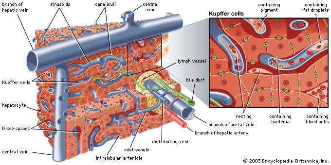
Microscopic structure of the liver. Liver cells, or hepatocytes, have direct access to the liver's blood supply through small capillaries called sinusoids. Hepatocytes carry out many metabolic functions, including the production of bile. Kupffer cells line the liver's vascular system; they play a role in blood formation and the destruction of cellular debris.
Hepatocytes occupy about 80 percent of the volume of the liver, and their cytoplasm (the area surrounding the nucleus) contains many mitochondria, which provide the energy needed for the many syntheticand metabolic functions of the liver cell. The cytoplasm also contains a series of long tubules, called the endoplasmic reticulum, which provides many enzymes essential to liver function. Some of the membranes of the endoplasmic reticulum appear granular, or rough, owing to the presence of ribosomes, which are responsible for forming specific polypeptide (protein) chains after having had the amino group removed (deamination) and having been converted into glucose through a process called gluconeogenesis. The ammonia released from gluconeogenesis is converted to urea in the hepatocyte by way of the urea cycle. The nonribosomal, or smooth, endoplasmic reticulum is where cytochromes (combinations of heme from hemoglobin with various proteins) and certain enzymes undertake the important hepatic functions of drug and hormonal metabolism and also cholesterol synthesis. Hepatocytes also conjugate with carbohydrate components of bilirubin and other fat-soluble metabolic and foreign compounds and thereby are made soluble in water. Bilirubin is the product of hemoglobin metabolism that is formed in the bone marrow and the lymphatic tissue and is carried to the liver after becoming bound to plasma albumin. It is released at the hepatocytic sinusoidal membrane and is transported to the smooth endoplasmic reticulum, where it is conjugated with one or two molecules of glucuronic acid and thereby becomes soluble in water and excretable in bile. The Golgi apparatus, a series of tubular structures between the endoplasmic reticulum and the canaliculus, acts as a transport station for newly made proteins and other hepatocytic products before they are conveyed to other parts of the cell or out of the cell entirely. Lysosomes, another important cytoplasmic constituent, are responsible for the intracellular storage of pigments, such as iron or copper, and for the digestion of certain contents, such as glycogen or foreign particles. The nucleus of the hepatocyte guides replication of the cell and transmits genetic material in the form of messenger ribonucleic acid (mRNA) from deoxyribonucleic acid (DNA) to organelles located in the cytoplasm.
The major functions of the liver are to participate in the metabolism of protein, carbohydrates, and fat; to synthesize cholesterol and bile acids; to initiate the formation of bile; to engage in the transport of bilirubin; to metabolize and transport certain drugs; and to control transport and storage of carbohydrates.
Sources:
https://www.ncbi.nlm.nih.gov/pubmedhealth/PMHT0022855/
Image Credits
shutterstock.com, webcache.googleusercontent.com, competitionaffairs.blogspot.in, sciencelearn.org.nz, pinterest.ie, slideplayer.com
https://www.consumerhealthdigest.com/health-conditions/gastrointestinal-tract.html
https://library.med.utah.edu/WebPath/HISTHTML/NORMAL/NORMAL10.html
http://pediatrics.aappublications.org/content/113/Supplement_3/1044 Pediatrics
https://patient.info/health/dyspepsia-indigestion/features/the-digestive-system Deseases
Unit 10
Nervous system

T opic Nervous System
Grammar Continuous Tense Revision Simple Tense, Degrees of Comparison of Adjectives and Adverbs
Vocabulary Nervous System, Noun suffix – on, - ance (- ence) Verb suffix - izePrefix inter - Revision Noun suffixes – um, - us Adjective suffix – al, - ic Verb suffix - ate
Reading ‘Nervous System’, ‘Cerebrospinal fluid’
Listening ‘Peripheral nervous system’ (video)
Speaking Pair work (Structure and Functions of NS), Group Work (Peripheral nervous system), Summary
Getting started