
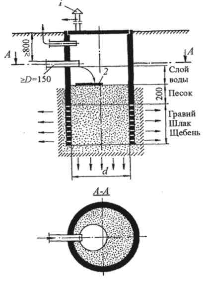
Индивидуальные очистные сооружения: К классу индивидуальных очистных сооружений относят сооружения, пропускная способность которых...
Индивидуальные и групповые автопоилки: для животных. Схемы и конструкции...
Индивидуальные очистные сооружения: К классу индивидуальных очистных сооружений относят сооружения, пропускная способность которых...
Индивидуальные и групповые автопоилки: для животных. Схемы и конструкции...
Топ:
Выпускная квалификационная работа: Основная часть ВКР, как правило, состоит из двух-трех глав, каждая из которых, в свою очередь...
Определение места расположения распределительного центра: Фирма реализует продукцию на рынках сбыта и имеет постоянных поставщиков в разных регионах. Увеличение объема продаж...
Методика измерений сопротивления растеканию тока анодного заземления: Анодный заземлитель (анод) – проводник, погруженный в электролитическую среду (грунт, раствор электролита) и подключенный к положительному...
Интересное:
Как мы говорим и как мы слушаем: общение можно сравнить с огромным зонтиком, под которым скрыто все...
Влияние предпринимательской среды на эффективное функционирование предприятия: Предпринимательская среда – это совокупность внешних и внутренних факторов, оказывающих влияние на функционирование фирмы...
Наиболее распространенные виды рака: Раковая опухоль — это самостоятельное новообразование, которое может возникнуть и от повышенного давления...
Дисциплины:
|
из
5.00
|
Заказать работу |
|
|
|
|
An electrocardiogram (e-lek-tro-KAR-de-o-gram), also called an EKG or ECG is a simple, painless test that records the heart’s electrical activity. To understand this test, it helps to understand how the heart works.
With each heartbeat, an electrical signal spreads from the top of the heart to the bottom. As it travels, the signal causes the heart to contract and pump blood. The process repeats with each new heartbeat.
The heart’s electrical signals set the rhythm of the heartbeat. From more detailed information and animations, go to the Health Topics How the Heart Works article.
An EKG shows:
Doctors use EKGs to detect and study many heart problems, such as heart attacks, arrhythmias (ah-RITH-me-ahs), and heart failure. The test’s result also can suggest other disorders that heart function.
Other Names for an Electrocardiogram
An electrocardiogram also is called an EKG or ECG. Sometimes the test is called a 12-lead EKG or 12-lead ECG. This is because the heart’s electrical activity most often is recorded from 12 different places on the body at the same time.
Who Needs an Electrocardiogram?
Your doctor recommend an electrocardiogram (EKG) if you have signs or symptoms that suggest a heart problem. Examples of such signs and symptoms include:
You may need to have more than one EKG so your doctor can diagnose certain heart conditions.
An EKG also may be done as part of a routine health exam. The test can screen for early heart disease that has no symptoms. Your doctor is more likely to look for early heart disease if your mother, father, brother, or sister had heart disease – especially early in life.
You may have an EKG so your doctor can check how well heart medicine or a medical device, such as a pacemaker, is working. The test also may be used for routine screening before major surgery.
Your doctor also may use EKG results to help plan your treatment for a heart condition.
What to Expect Before an Electrocardiogram
You don’t need to take any special steps before having an electrocardiogram (EKG). However, tell your doctor or his or her staff about the medicines you’re taking. Some medicines can affect EKG results.
What to Expect During an Electrocardiogram
An electrocardiogram (EKG) is planes and harmless. A nurse or technician will attach soft, sticky patches called electrodes to the skin of your chest, arms, and legs. The patches are about the size of a quarter.
|
|
Often, 12 patches are attached to your body. This helps detect your heart’s electrical activity from many areas at the same time. The nurse may have to shave areas of your skin to help the patches stick.
After the patches are placed on your skin, you’ll lie still on a table while the patches detect your heart’s electrical signals. A machine will record these signals on graph paper or display them on a screen.
The entire test will take about 10 minutes.
Answer the questions:
5. How many minutes will it take you to the entire test?
Radiological Researches
The x-ray method of research of a thorax is used for recognition, first of all, diseases of lungs – pneumonia, tuberculosis, tumors, professional defeats, and also for diagnostics of heart diseases, diseases of cardiac muscle, pericardium diseases. The method helps with recognition of changes of a backbone, lymph nodes. The x-ray method for routine inspections is widely used, in particular at identification of early symptoms of tuberculosis, tumors, professional diseases when other symptoms of these diseases still are absent. X-ray of abdominal organs carry out at a clinical picture of “a sharp stomach”, that is in the presence of complains of the patient to severe pains in a stomach. Usually thus diagnose the following diseases:
Cholecystitis, cholelithic illness
Pancreatitis
Abscesses and inflammatory diseases of abdominal organs
Gut ischemia
Acute appendicitis
Radiological research of bones and joints is carried out for recognition of the main diseases of joints – changes, tumors, inflammatory and dystrophic damage of joints. X-ray pictures of bones and joins are made in radiological offices. In drawing x-ray picture of a knee joint.Bones detain X-rays, they are well visible in pictures therefore any changes of bones – changes, cracks, reduction of the content of calcium, tumoral educations – are distinguished by means of radiological research. In pictures of joints their bone surfaces are well visible, a state the hryashchevykh of educations judge on width and a form of an articulate crack.
Radiological research of kidneys and urinary ways includes a survey X-ray analysis of a stomach when the picture, and research of urinary ways by means of contrast substances is simply made. It allows to receive the image of kidneys and urinary ways in X-ray pictures. Research objective – to receive approximate idea of an arrangement and size of kidneys, to find stones in uric ways. It is carried out in a X-ray office. The patient undresses, bares a stomach, lays down on a table of the X-ray device and pictures are made.
Radiological research of bodies of the gastrointestinal highway – the research methods, allowing to receive the image of these bodies on the screen of the X-ray device, and also to make pictures on a X-ray film.
|
|
Radiological research is applied to identification of diseases of a gullet (developmental anomalies, ulcers, tumors, narrowing of a gleam, diverticulums); stomach (ulcers, tumors), intestines (inflammatory diseases, tumors, diverticulums), and also violations of motive function of these bodies.
1. Allow- позволять
2. Arrangement - расположение
3. Approximately - ориентировочно
4. To apply- применять
5. Backbone - позвоночник
6. Bare- обнажать
7. Body – здоровый орган
8. Carry out - выполнять
9. To conduct – проводить исследование
10. Crack - трещина
11. Articulate crack – суставная щель
12. Screen – экран
13. Drawing X-ray picture – нарисункеснимок
14. Detain – задерживать (лучи)
15. Distinguish - распознавать
16. Developmental anomalies – порок развития
17. Education - образование
18. Image- изображение
19. Inflammation - воспаление
20. Identification - выявление
21. Tumor - опухоль
22. Thorax- грудная клетка
23. Research - исследование
24. Reduction - уменьшение
25. Recognition - распознать
26. Severe - острый
27. Stomach - желудок
28. Survey - обширный
29. Substance - вещество
30. Unpleasant - неприятный
31. Violation - двигательная функция
32. Office - кабинет
33. X-ray device – рентген аппарат
34. Make – производить снимки
35. X-ray film – рентгенологическая пленка
Answer the questions:
1. What does the thorax X-ray allow to receive?
2. Is the X-ray method of research of a thorax used for recognition of lungs diseases?
3. Where is the X-ray research of a thorax made?
4. Does the patient feel any unpleasant feelings?
5. Name the main diseases of bones and joints?
6. What allows to receive the image of kidneys and urinary ways in X-ray pictures?
|
|
|

Таксономические единицы (категории) растений: Каждая система классификации состоит из определённых соподчиненных друг другу...
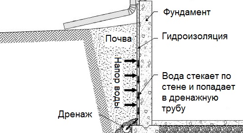
Общие условия выбора системы дренажа: Система дренажа выбирается в зависимости от характера защищаемого...
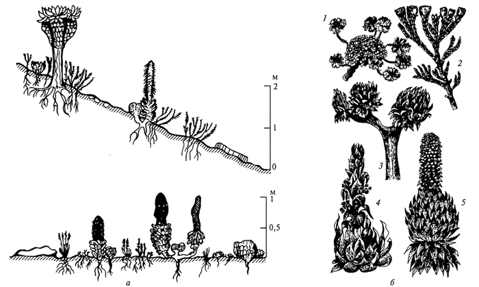
Адаптации растений и животных к жизни в горах: Большое значение для жизни организмов в горах имеют степень расчленения, крутизна и экспозиционные различия склонов...

Эмиссия газов от очистных сооружений канализации: В последние годы внимание мирового сообщества сосредоточено на экологических проблемах...
© cyberpedia.su 2017-2024 - Не является автором материалов. Исключительное право сохранено за автором текста.
Если вы не хотите, чтобы данный материал был у нас на сайте, перейдите по ссылке: Нарушение авторских прав. Мы поможем в написании вашей работы!