4. Translate the sentences paying attention to the words "one - ones" and "that - those":
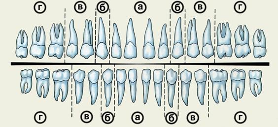
1. The upper second premolars have one root and the first ones have two roots.
2. The bones of the skull are those which compose the head and the face.
3. This lecture is more interesting than the one I attended last week.
4. The milk teeth are twenty in number and the permanent ones are thirty-two.
5. First baby teeth erupt at the age of 6 months after birth and those of the permanent set normally erupt at the age of 6 years.
6. Teeth of the first dentition must be treated as those of the second one.
Summary
Make a summary considering the stages of teeth development.
Tooth development
Before a human embryo is 3 weeks old, the stomodeum has been established. The primitive mouth is lined with ectoderm, beneath which is mesenchyme. The ectoderm gives rise to the oral epithelium, and the mesenchyme becomes the underlying connective tissue.
Odontogenesis is the name given to the origin and tissue formation of the teeth. Not all teeth start development at the same time. A tooth is formed from ectoderm and ectomesenchyme. Ectomesenchyme is derived from neural crest cells. Development begins with the formation of the primary dental lamina, extending along the jaws on a line where the teeth will later appear.
Concurrently with the development of primary dental lamina, at 10 places in the mandibular and at 10 places in the maxillary arch some cells of the dental lamina multiply and 10 little knobs of epithelial cells are formed on the dental lamina in each jaw. Each of these knob-shaped structures is an early enamel organ. It is the beginning of the tooth germ of a primary tooth. A tooth germ is derived from two embryonic tissues: the part that develops from the dental lamina originate from ectoderm and the remaining parts originate from mesenchyme underlining ectoderm.
As the dental lamina enlarges, the enamel organ acquires the shape of a cap. By the 8th week in utero this cap formation is seen in the enamel organs of the deciduous incisor tooth germs. The connective tissue inside the cap undergoes a number of changes and becomes the dental papilla. The connective tissue beneath the dental papilla becomes fibrous and encircles the papilla forming the dental sac.
The crown and the root of the tooth grow as the result of the deposition of new layers of enamel and dentin on previously formed layers. When the final size of the enamel crown has been attained, the enamel-forming cells disappear and further formation of enamel is impossible.
The fetus derives its mineral from mother through placenta. That’s why the dentin formed in utero is always of a more homogenous texture and more mineralized than that laid down postnatally. In the young child, the pulp cavity is quite large relative to amount of dentin already formed. For this reason, capping of teeth may be delayed for several years until the formation of the dentin has been completed. So the infant is fully dependent on the mineral obtained through ingestion.
Translation
The Teeth
A tooth may be divided into crown and root, the crown being covered by enamel and the root by cementum. The two surfaces meet at the cement-enamel junction which is visible as the cervical line on the neck of the tooth. In the healthy mouth of a young adult, the level of gingival attachment will be coronal to the cervical line. The anatomical crown ends at the cervical line. Variations in clinical crown length are often produced by different levels of gingival attachment to the tooth and are seen in patients suffering from gingival recession and hence showing increased clinical crown length. Short clinical crowns are seen in the teeth that have worn excessively, commonly due to bruxism, or where teeth have been worn down by attrition.
Incisors and canines have four axial surfaces converging in an incisal edge. Premolars and molars have five surfaces, the incisal edge being replaced by an occlusal surface. The surface of the crown shows many elevations and depressions which make up the typical appearance of the tooth. The following terms are used in the description of crown anatomy:
Cusps: an elevation or mound on the occlusal surface.
Cingulum: the lingual convex bulge on an anterior tooth.
Tubercle: a small elevation on some part of the crown produced by an extra formation of enamel and dentine. These are quite frequently seen buccally on deciduous first molars (the tubercle of Zuckerkandl) and lingually on upper first molars (the cusp of Carabelli).
Ridge: a linear elevation on the surface of a tooth. A good example is the marginal ridge found on the mesial and distal surfaces of molars and premolars.
Fissure: an irregular linear depression in the tooth surface. A pit is a small pinpoint depression.
Developmental groove: a developmental deformity in the crown and/or root of a tooth. This type of defect will encourage the formation of a periodontal pocket, particularly when it involves the root, because dental plaque will collect there undisturbed. Grooves are sometimes seen on permanent upper lateral incisors, especially on palatal surfaces.
Mamelon: any one of the three rounded protuberances found on the incisal edges of recently erupted anterior teeth. Mamelons wear away quickly, usually within two years of eruption.
Facet: a small, smooth, flat surface seen on the occlusal aspect of the crown indicating an abnormal pattern of wear on the enamel.
Perikymata: seen commonly on recently erupted incisors as a series of horizontal redges running parallel to the incisal edge and quite often affecting the whole of the labial surface of the crown.
Speaking